|
|
||
05/06/05 |
|
Anatomy 101 revisited! You'll find it helpful to have a basic knowledge of muscle anatomy before you head to the gym. Here are common and technical descriptions of key muscles. |
||
|
|
Chest
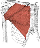
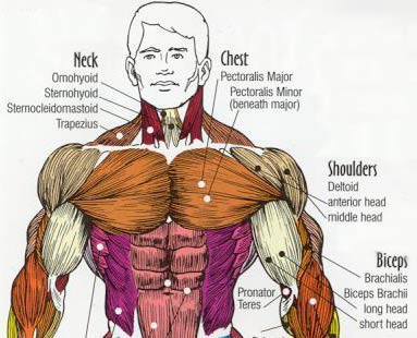
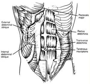
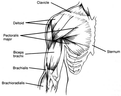
Simple Description Common Name: Chest muscles Location on the Body: Place your hand on your chest over your heart. Motion Performed: When all fibers work together, they bring your arms across your body. Visualize wrapping your arms around a big tree. Technical Description Scientific Name: Pectoralis major Location on the Body: The pectoralis muscle originates at the clavicle, sternum, cartilage of ribs 1-6, and inserts on the lateral aspect of the anterior humerus. Motion Performed: The primary motion of concern is shoulder horizontal adduction.
Abdomen
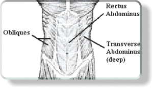
Simple Description Common Name: Abs Location on the Body: The Abdominal muscles sit on the front and sides of the lower half of the torso, originating along the rib cage and attaching along the pelvis. Motion Performed: Keeps your insides in. Technical Description Scientific Name: The Abdominals are composed of several muscles: the Rectus Abdominus, Transverse Abdominus, and the External and Internal Obliques. Location on the Body: The Abdominal muscles sit on the front and sides of the lower half of the torso, originating along the rib cage and attaching along the pelvis. Motion Performed: Rectus Abdominus: Flex the spine (bringing the rib cage closer to the pelvis). This is seen in the abdominal crunching movement. Transverse Abdominus: Acts as a natural weight belt, keeping your insides in. This muscle is essential for trunk stability. Internal and External Obliques: Work to rotate the torso and stabilize the abdomen.
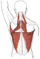
Back
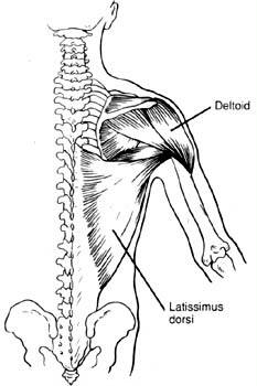
Simple Description Common Name: Lats Location on the Body: The lats run below the shoulders to your lower back (your belt line). To locate the lats, take your left hand and reach behind your right armpit where the "wide" part of the lats is located. Motion Performed: Envision pulling a door open (shoulder extension) or grabbing overhead and pulling up on a chin-up bar (shoulder adduction). Technical Description Scientific Name: Latissimus dorsi Location on Body: The lats originate from T-6 down to the sacrum. The fibers then wrap around, between the rib cage and the arm, and converge at a point on the anterior humerus (next to the chest). Motion Performed: The lats perform shoulder extension as well as shoulder adduction against resistance.
Shoulders
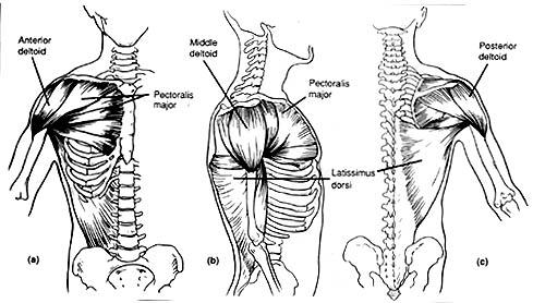
Simple Description Common Name: Shoulder muscles Location on the Body: The shoulders are the muscles underneath a football player’s shoulder pads. Motion Performed: The shoulders move your upper arm up, back and to the side. Technical Description Scientific Name: Deltoids Location on the Body: The deltoid runs between the spine and the clavicle of the shoulder girdle to the deltoid tuberosity of the humerus and has three distinct parts: anterior, middle and posterior. Motion Performed: Shoulder flexion is performed by the anterior deltoid and anterior portion of the middle deltoid. Horizontal abduction is best performed by the posterior deltoid and the posterior portion of the middle deltoid. Together, these two motions will sufficiently work the whole deltoid complex.
Biceps
Simple Description Common Name: Biceps Location on the Body: The biceps are the muscles in front of your arms. Motion Performed: When you bend your arm to pick something up, you use your biceps. Technical Description Scientific Name: Biceps brachii Location on the Body: The short or medial head attaches to the coracoid process of the scapula; the long or lateral head attaches to the glenoid. Both run to a tendon that travels between the radius and ulna attaching medially. Motion Performed: Flexes elbow joint and supinates forearm. Although the bicep crosses the shoulder joint, it is a better shoulder-joint stabilizer than mover.
Triceps
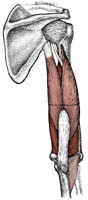
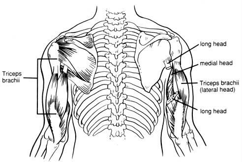
Simple Description Common Name: Triceps Location on the Body: The triceps are on the back of the upper arm. Motion performed: The triceps straighten your elbows when you push yourself out of a chair. Technical Description Scientific Name: Triceps brachii Location on the Body: The long head attaches to the infraglenoid tubercle of the scapula; the lateral head and medial head attach to the posterior humerus. All insert to a common tendon into the olecranon process of the ulna. Motion Performed: Powerful forearm extensor. Longhead tendon may help stabilize shoulder joint and assist in arm adduction.
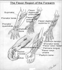
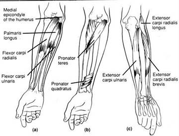
Forearm
Simple Description Common Name: Forearms Location on the Body: Lower arms, above the hands. Motion Performed: Extend and flex the wrist. Technical Description Scientific Name: Flexor muscles and Extensor muscles. Location on the Body: The muscles originate close to the elbow and extend all the way to the tips of the fingers. Motion Performed: Flexor muscles: Responsible for curling the fingers palm ward and bending the wrists palm ward. Extensor muscles: Responsible for bending the wrists toward the back of the hand.
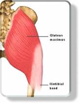
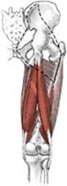
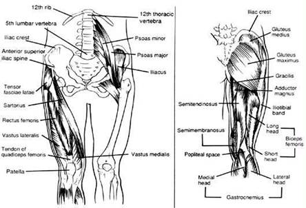
Glutes
Simple Description Common Name: Glutes Location on the Body: The butt. Motion Performed: Moving the leg back and outward Technical Description Scientific Name: Gluteus Maximus, Gluteus Medius, Gluteus Minimus and Iliotibial Band Location on the Body: Gluteus Maximus originates along the pelvic bone crests and attaches to the rear of the femur. Gluteus Medius and Minimus originate in the same spot as the Maximus but attach to the side of the femur. The Iliotibail Band is made only of connective tissue. Motion Performed: Gluteus Maximus: Hip extension (moving the thigh to the rear). Gluteus Medius and Minimus: Serve to abduct (move away from the centerline of the body) the leg. Iliotibial Band: Serves to transfer the force of abduction (moving the leg away from the centerline of the body) to the leg.
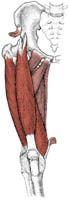
Quads
Simple Description Common Name: Quads Location on the Body: To locate the four quad muscles, place both your hands on the front of the thighs. Motion Performed: The quads straighten the knee. Quads are used every time you take a step. Technical Description Scientific Name: Quadriceps femoris (rectus femoris, vastus intermedius, vastus medialis, vastus lateralis) Location on the Body: The quadriceps are composed of four individual but interdependent muscles. The vastus lateralis and medialis attach to the posterior aspect of the femur, laterally and medially respectively. The intermedius attaches to the femur anteriorly. They converge to the tibial tuberosity. Motion Performed: The quadriceps is a powerful knee extensor. In addition, the two joint muscles act as knee extensors thigh flexors at the hip.
Hamstrings
Simple Description Common Name: Hams Location on the Body: The hamstrings are the muscles on the back of your leg. Motion Performed: The hams bend the knee. For example, they lift your leg as you walk. Technical Description Scientific Name: Hamstrings (biceps femoris, semitendinosus, semimembranosus) Location on the Body: The hamstrings are a group of three muscles. The semimembranosus and semitendinosus both originate on the ischial tuberosity and insert on the medial tibia. The bicep femoris has two heads. The long head originates on the ischial tuberosity and the short head on the femur. They both insert on the head of the fibula via a common tendon. Motion Performed: Extends thigh and flexes knee; the biceps femoris laterally rotates leg, especially when knee is flexed. The semitendinosus and semimembranosus medially rotate the leg.
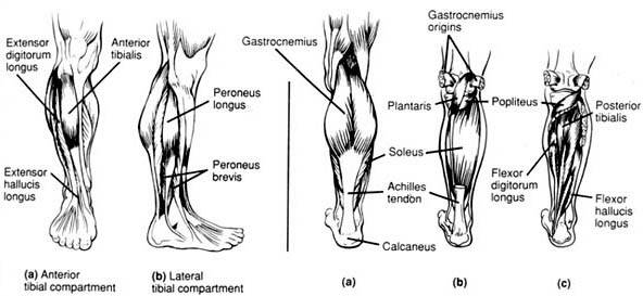
Calves
Simple Description Common Name: Calves Location on the Body: The calves are the muscles in the back of the leg that lie underneath your tube socks (assuming you pull them up high). Motion Performed: The calves are the muscles that allow you to reach high on your toes to reach into a cabinet and also stabilize your ankles when you walk. Technical Description Scientific Name: Triceps surae (gastrocnemius and soleus) Location on the Body: Gastrocnemius. The lateral head attaches to the lateral femur and the medial head attaches to the medial femur. Both heads attach to the calcaneous via a common tendon. The soleus attaches to the tibia, fibula and the calcaneous. Motion Performed: Gastrocnemius—the plantar flexes the foot when the knee is extended. Because it also crosses the knee joint, it can assist in flexion of the knee when the foot is dorsiflexed. Soleus—the plantar flex foot also is an important locomotor and postural muscle during walking and running.
|
|
This site was last updated 01/26/05